Galería de imágenes obtenidas con el microscopio confocal
|
|
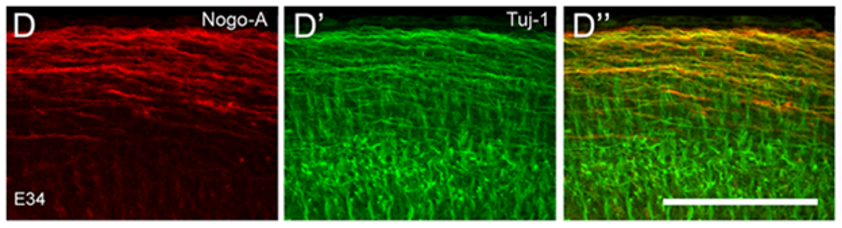
Figs D – Nogo-A is prominent in axons of the retinorecipient layers of the lizard embryos. Counterstain with Tuj-1 and signal overlay in D – IP. Dra. M. Monzón Mayor
|
|
|
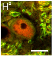
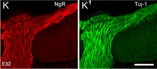
Fig H2 – PLP oligodendrocytes display Nogo-A signal enriched in the cytoplasm of the adult lizard; Figs. K. K1 – Tuj-1+ RGC axons show distinct NgR labeling in the retinal NFL – Coordinadora IP. Dra. M. Monzón Mayor. Grupo Neurogliociencia
|
|
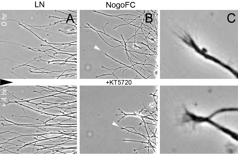
Time-lapse analysis of the effect of the pkA inhibitor KT5720 on lizard RGC axon growth in vitro. J. Comp. Neurol. 525:936–954,2017 – Coordinadora IP. Dra. M. Monzón Mayor – Grupo de Neurogliociencia
|
|
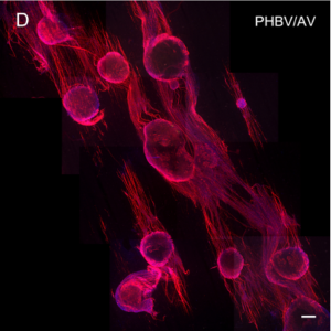
Immunofluorescence staining of rat DRG explants cultures with beta-III tubulin. Aligned neurite outgrowth directed by the underling matrix of hybrid PHBV/AV explant – Coordinadora IP: Dra. M. Monzón Mayor – Grupo de Neurogliociencia